Adobe PDF
(42.34 MB)
Brochure
Download
| Title Details: | |
|
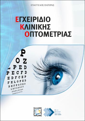
Handbook of Clinical Optometry |
|
| Authors: |
Pateras, Evangelos |
| Subject: | NATURAL SCIENCES AND AGRICULTURAL SCIENCES > PHYSICS > > MEDICINE AND HEALTH SCIENCES, LIFE SCIENCES, BIOLOGICAL SCIENCES > HEALTH SCIENCES > MEDICAL ILLUSTRATION MEDICINE AND HEALTH SCIENCES, LIFE SCIENCES, BIOLOGICAL SCIENCES > HEALTH SCIENCES > MEDICINE > SURGICAL SPECIALTIES > OPHTHALMOLOGY MEDICINE AND HEALTH SCIENCES, LIFE SCIENCES, BIOLOGICAL SCIENCES > HEALTH SCIENCES > OPTOMETRY |
| Keywords: |
Eye
Optometry Eye Investigation and Examination Techniques Clinical eye refraction Corneal topography Optical coherence tomography of the eye Slit lamp Ocular biometry Ophthalmoscopy Perimetry, Visual fields Tonometry Confocal and Specular microscopy of the eye Clinical visual electrophysiology OCT angiography Color vision Visual acuity Pediatric optometry Binocular vision Retinoscopy Contrast sensitivity testing |
| Description: | |
| Abstract: |
The Handbook of Clinical Optometry has been designed to facilitate undergraduate and postgraduate students in Optics & Optometry. The aim is to present the latest developments in eye imaging equipment and the steps to be followed for a complete optometric examination. Specifically, it includes the protocol for a complete optometric examination which presents the objective examination of the refractive state of a patient through retinoscopy or automated refraction. It also analyzes the procedure of subjective refraction using a simple trial frame and trial lenses or using a phoropter. The slit lamp is presented, and its techniques for assessing the physiology of the eye and the corneal topography is analyzed with the presentation of modern topographers and its use in the detection of keratoconus. Reference is made to special imaging techniques such as optical coherence tomography (AS-OCT / POST-OCT), and modern OCT angiography. Imaging techniques also include ocular biometry, A-scan, B-scan ultrasound, UBM - IOL-Master, confocal and specular microscopy, direct and indirect ophthalmoscopy and the use of a non-mydriatic camera for retina observation. The perimetry technique used to evaluate the visual fields is described and a comparison is made between known perimeters as well as evaluation of their findings. The protocol also includes contrast sensitivity testing, color vision testing with the corresponding diagnostic tests, tonometry, gonioscopy, electrophysiological methods of vision examination with EOG, ERG, and VEP evoked potentials and finally reference is made to stereoscopic binocular vision and its testing techniques. Finally, all the necessary diagnostic tests are presented in a preoperative examination before eye surgeries and a small report on pediatric optometry. The handbook is completed with basic knowledge of ophthalmic lenses, such as aspheric, multifocal and prism.
|
| Linguistic Editors: |
Ελένη, Helen |
| Graphic Editors: |
Σταύρος, Stavros |
| Type: |
Laboratory Guide |
| Creation Date: | 20-12-2021 |
| Item Details: | |
| ISBN |
978-618-85370-7-1 |
| License: |
Attribution - NonCommercial - ShareAlike 4.0 International (CC BY-NC-SA 4.0) |
| Handle | http://hdl.handle.net/11419/8014 |
| Bibliographic Reference: | Pateras, E. (2021). Handbook of Clinical Optometry [Laboratory Guide]. Kallipos, Open Academic Editions. https://hdl.handle.net/11419/8014 |
| Language: |
Greek |
| Publication Origin: |
Kallipos, Open Academic Editions |


